
Laser Bioanalytics is a Colorado-based life sciences instrumentation company dedicated to advancing the frontiers of spatial biology and structural proteomics. Originating from 25 years of mass spectrometry research, we engineer next-generation platforms that integrate high-precision deep-ultraviolet and infrared lasers with targeted in situ photochemistry and precise laser ablation microsampling. Our cutting-edge workflows empower researchers to bypass traditional analytical limits thereby enabling the spatial mapping of lipid isomers, the artifact-free extraction of native-state biomolecules, and the high-resolution structural profiling of folded proteins directly within the tissue microenvironment.
Deep Ultraviolet Laser Ablation Microsampling
Soft protein extraction: deep-uv cold ablation for native-state proteomics
Application: Precision microsampling, structural proteomics, genomics
This system utilizes a deep-ultraviolet laser to execute cold ablation, enabling high-precision physical removal of targeted tissue regions. It extracts intact, chemically unaltered biomolecules from precise spatial coordinates for downstream, off-line characterization. By minimizing thermal damage and chemical artifacts it preserves the native states of fragile biomolecules for downstream analysis.

Currently commercial laser ablation microsampling instruments use two methods for tissue region excision: laser cutting and tissue adhesion for laser microdissection. Laser cutting microdissection uses a focused high energy laser to obliterate tissue at the perimeter of a tissue region of interest to separate a small solid mass of tissue from its surroundings. The isolated piece is separated from the bulk by an additional laser pulse or film adhesion. The high laser energy used for cutting can damage the tissue adjacent to the cut line and lysis and additional separation steps are required for extraction of biomolecules from the intact tissue pieces.
Adhesion laser microdissection methods typically use laser melting which involves heating that can disrupt weak biomolecule interactions. Furthermore, adhesion of the tissue components to the polymer film can limit the extraction efficiency.
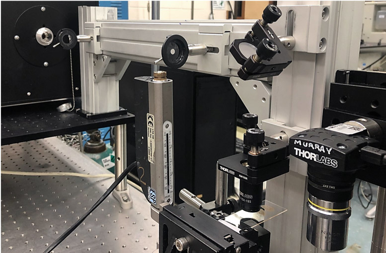
The Laser Bioanalytics deep ultraviolet laser ablaton microsampling system (DUV-LAM) uses the “cold” deep UV ablation method and irradiates the tissue spot by spot to disrupt the structure and eject the tissue components in the plume of ejected particulate material. The collected biomolecules are efficiently extracted and do not experience that fragmentation or disruption of weak interactions that can occur through high energy cutting, heating, and lysis.
References
- Murray, K., Donnarumma, F., & Lawai, O. (2021). Devices and Methods For Deep UV Laser Ablation.
Photochemical MALDI
Breaking the isobaric barrier: spatial mapping lipid double-bonds
Application: Double bond localization in MALDI lipid imaging
Matrix-assisted laser desorption/ionization (MALDI) is a soft ionization technique used in mass spectrometry to analyze fragile, high-molecular-weight molecules, such as proteins, peptides, and synthetic polymers, with minimal fragmentation. MALDI imaging uses this method to map the spatial distribution of biomolecules in thin tissue sections.

This project is aimed at developing a benchtop instrument for photochemical modification of tissue prior to MALDI imaging. This technology photochemically modifies unsaturated lipids within thin tissue sections via targeted photochemical reactions. By pinpointing the locations of aliphatic carbon-carbon double bonds, it maps lipid isomers directly from the tissue regions. This workflow generates diagnostic fragments that successfully overcome the limitations of standard isobaric mass spectrometry imaging.
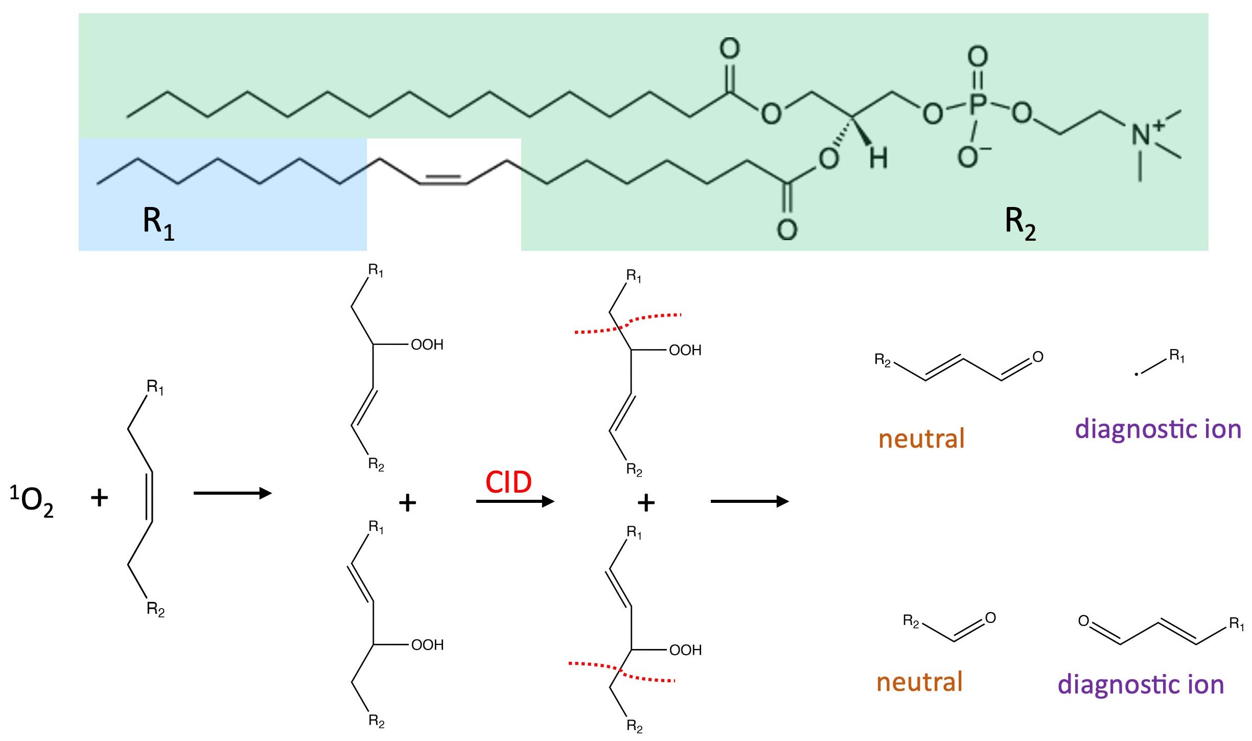
of a photosensitizer and the reaction products lead
to diagnostic ion formation in tandem mass spectrometry.
Pulsed laser irradiation of singlet oxygen phosensitizer creates hydroperoxide products that help pinpoint double bond location in unsaturated lipids using tandem mass spectrometry.
This project is funded by the National Institutes of Health project number 1R43GM156259-01, “Photochemical Tissue Modification for MALDI Mass Spectrometry Imaging”
Photochemical Laser Ablation Microsampling
In situ architecture: mapping secondary protein structure via photochemical oxidation
Application: Structural proteomics, lipidomics
This approach drives in situ photochemical oxidation of localized tissue proteins using an ultraviolet laser combined with infrared laser ablation microsampling. The method probes solvent-accessible surfaces at micro-scale spatial resolution to identify and map the secondary structures of proteins and protein complexes.
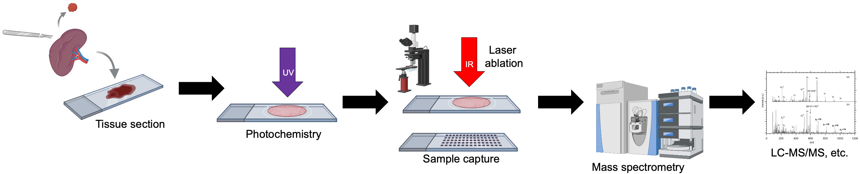
Laser microsampling is used to isolate specific, pure populations of cells from a tissue sections. By physically separating cells of interest from surrounding normal or stromal tissue, researchers can perform high-purity molecular analysis (DNA, RNA, or protein) that would otherwise be diluted by unwanted cell types. This Laser Bioanalytics system combines laser photochemistry with laser microsampling.
The photochemical laser ablation microsampling (PC-LAM) system uses a novel infrared laser ablation microsampling microscope for fast and efficient selection and collection of biomolecules from tissue sections. A pulsed ultraviolet laser is used to initiate photochemical reactions in tissue sections for selective labeling of biomolecules. The reactions are rapidly quenched, and the modified biomolecules processed to obtain structural information.
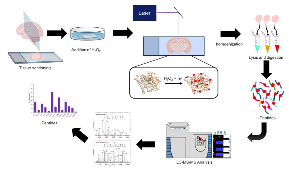
Deep Ultraviolet Laser Protein Footprinting
Structural topology unlocked: deep-uv footprinting of proteins and protein complexes
This structural biology technique uses deep ultraviolet laser irradiation to photochemically label solvent-exposed amino acid side chains. It maps the higher-order structure and structural topology of folded proteins under native-like conditions. Pairing high-speed modification with bottom-up proteomics workflows systematically reveals protein folding, tertiary topologies, and binding interactions.

References
- Lawal, et al. J. Am. Soc. Mass. Spectrom. 2019, 30, 2196.